News and Views by: Gabe Jones (gajones@davidson.edu)
Primary Article: Oikonomou et al. 2020
This web page was produced as an assignment for an undergraduate course at Davidson College.
Join me on a journey through the genome where novel vivo mRNA display reveals the orchestrated dance between genetic transcripts and protein phenotypes. By use of this molecular tagging system, this new frontier of next-generation sequencing identifies proteins in various vivo functional assays through the identification of its nucleic acid sequence.
There is an intricate orchestra of life within our cells, where a fundamental process unfolds governed by a series of sequences that act as the messenger of genetic information: mRNA, or messenger RNA. At the heart of this molecular choreography lies the extraordinary journey from DNA to proteins, thanks to mRNA. Think of DNA as the blueprint, housing the instructions for every protein our cells need. But it’s mRNA that ventures forth from the cell’s nucleus, carrying the genetic blueprints to the protein factories known as ribosomes. This pivotal role of mRNA in protein synthesis forms the very foundation of proteomics, the study of proteins and their functions within biological systems.
Classical proteomics typically involves the use of Mass-Spectrometry, a technique that requires complicated sample preparation, protein extraction, and separation of proteins. The precise identification of individual protein molecules is then based on computer algorithms deducing their mass and charge and comparing them to DNA and protein databases (Graves et al. 2002). While powerful, this method has limitations in scalability, cost, and the ability to capture proteins in their native live-cellular contexts. It is also not recommended for the use of an organism’s full protein display, otherwise known as the proteome. The authors of this paper sought to revolutionize proteomics by circumventing many of these described complications.
Remedying most all classical issues, the advent of vivo mRNA display marks a significant leap forward providing standardization, scalability, and cost-efficiency to proteome characterization. This innovative technology bridges the gap between genotype and phenotype by linking in vivo expressed proteins with their encoding mRNAs via a stem-loop RNA binding domain interaction (Oikonomou et al. 2020). By harnessing the power of next-generation DNA sequencing, in vivo mRNA display offers a promising avenue for comprehensive proteomic analysis within native cellular contexts.
The authors accomplished this feat by using the MS2 bacteriophage coat protein and its cognate RNA stem loop to create a stable linkage of proteins to their encoding mRNAs in vivo. MS2 has been thoroughly investigated in the past to deduce its RNA binding mechanism and utilized as a reporter system to track mRNA in living cells (Peabody 1993) Here, they modified this tagging system by fusing MS2 to a target protein with the cognate stem loop sequence placed downstream of the protein’s gene, creating a direct, detectable, association between the proteins and mRNA in vivo. In their initial testing they confirmed that this methodology has no adverse effects on wild-type versus mutant protein expression although some proteins are unable to display their mRNAs effectively. For example, as the cells go about expressing the protein in their natural context the abundance of mRNA can be compared before and after an assay to measure the production under different simulated biological conditions.
To confirm the success of their tagging system, the researchers conducted a series of experiments. They first demonstrated the stable interaction between the translated proteins and their encoding mRNAs through immunoprecipitation assays using magnetic beads specific for the detection of MS2 to target protein fusion constructs. They observed a significant enrichment of the target proteins mRNA which confirmed that when the protein is synthesized within the cell, it binds to its own mRNA via the MS2 coat protein and the RNA stem loop (Figure 1). Further confirmation was accomplished via competitive assays by introducing two different sets of genetic constructs into yeast cells, each labeled with in vivo display tags. Despite the mixed population of cells that resulted from their growth, the researchers were still able to accurately measure the quantity of each protein produced via mRNA sequencing.
Through a combination of genetic engineering and high-throughput sequencing, the novel method showcases its potential to revolutionize proteomic research by providing a straightforward cost-effective solution for large scale protein analysis. While this approach is still dependent on the construction of dense DNA and protein libraries, the authors argue that the initial investment would pay-off overtime as this resource is applicable to diverse plethora of proteomic assays. I agree with their sentiment as a single mass spectrometry experiment can cost over $1,000 when the same protein samples can be generated and processed for roughly a tenth of the cost with vivo mRNA display. Getting this technology across the globe could revolutionize the way proteomics is carried out especially in lower to middle class countries where it may not be feasible to spend hundreds of thousands of dollars just running mass spectrometry on protein samples that may not behave the same as they do in live cells.
The authors and I both carry a vision that requires future refinement of the technique but the hope that its application exists with a glass ceiling ready to be applied in ways we cannot think of today. As I mentioned earlier not every protein folded and or bound to the mRNA as anticipated, while most of the tested proteins did, further investigation is needed into the thermodynamics of protein formation/binding in these exceptions. Despite these challenges, the promise of in vivo mRNA display technology is profound. With continued improvement, its wide application holds the potential to optimize peptides and proteins for therapeutic benefits alike in vitro phage display did in the past (Clackson et al. 1991). This advancement offers hope for addressing medical needs but also raises ethical considerations regarding the equitable use of such powerful technologies that ever so quickly nowadays get relegated only to the highest, most wealthy, classes. As researchers navigate these complexities, they, and I, both are filled by the optimism that this research will pave the way for transformative breakthroughs in biomedicine through the application of proteomics.
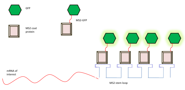
Figure 1) Schematic of MS2 bound GFP binding to mRNA. This figure provides a visual example of a target protein, GFP, and how it would bind to its own mRNA given the use the MS2 in vivo mRNA display technique. (Kumar 2023)
- Peabody DS. The RNA binding site of bacteriophage MS2 coat protein. The EMBO Journal 1993; 12: 595. [PMID: 8440248 DOI: 10.1002/j.1460-2075.1993.tb05691.x]
- P O, R S, S T. In vivo mRNA display enables large-scale proteomics by next generation sequencing. Proceedings of the National Academy of Sciences of the United States of America 2020; 117. [PMID: 33037152 DOI: 10.1073/pnas.2002650117]
- Kumar P. RNA Imaging. Materials and Methods (e-pub ahead of print 19 February 2023).
- Graves PR, Haystead TAJ. Molecular Biologist’s Guide to Proteomics. Microbiology and Molecular Biology Reviews 2002; 66: 39. [PMID: 11875127 DOI: 10.1128/MMBR.66.1.39-63.2002]
- Clackson T, Hoogenboom HR, Griffiths AD, Winter G. Making antibody fragments using phage display libraries. Nature 1991; 352: 624–628. [DOI: 10.1038/352624a0]
© Copyright 2022 Department of Biology, Davidson College, Davidson, NC 28036.

0 thoughts on “Unveiling a Genetic Symphony: mRNA the Maestro of Novel Proteomic Discoveries”