Disclaimer: This web page was produced as an assignment for an undergraduate course at Davidson College.
A comprehensive genome-wide atlas of epigenetic profiles in gene regulatory regions unveils differences between healthy and adaptive kidney cells, shedding light on potential therapeutic targets for kidney repair.
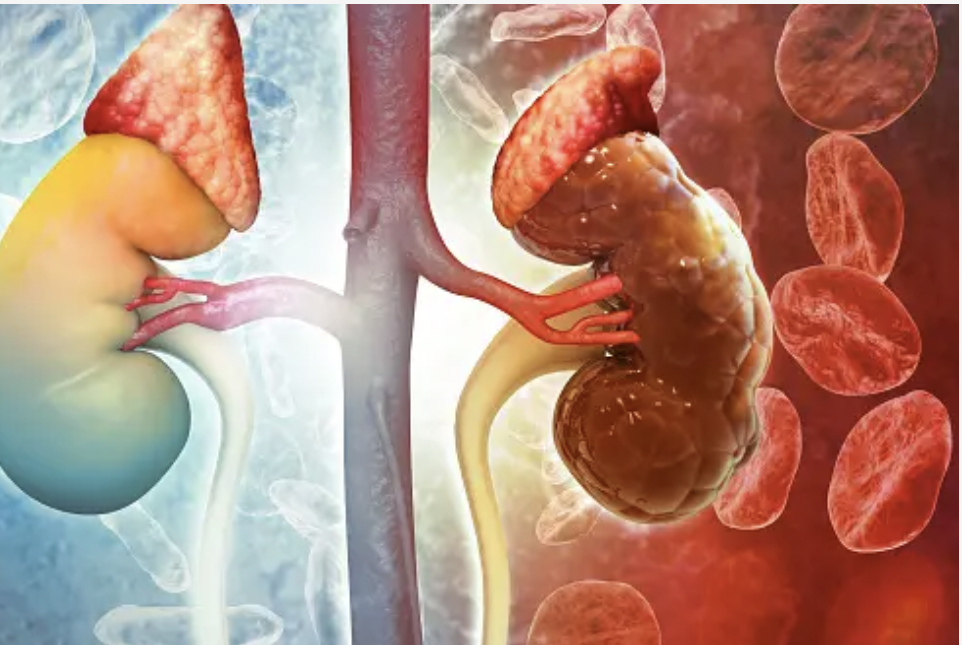
Kidney cells exhibit gene expression changes after being exposed to damage such as infection and environmental toxins. Previous studies illustrated the gene expression changes in the adaptive states (after injury) of different types of kidney cells such as proximal tubule (PT) and the thick ascending limb (TAL) epithelial cells.1 However, how epigenetic regulation affects the transition of gene expression from healthy to damaged kidney cells is poorly understood. Epigenetic markers are chemical compounds that modify DNA and alter gene expression without changing the DNA sequence. DNA methylation and histone modification are the most common epigenetic regulators that have significance in kidney diseases.2 DNA methylation occurs when a methyl group(-CH3) group is added to a cytosine base in our DNA sequence, whereas histone modification is a post-translational alteration of histone proteins, to whom DNA wraps around to make a nucleosome. The understanding of the roles of these epigenetic regulations in different kidney cells is vital to elucidating the gene expression pattern and chromatin landscape in normal and adaptive states.
Image courtesy of Istockphoto.com
Gisch et al. developed a comprehensive atlas of genome-wide epigenomic features of the kidney.3 They first identified open chromatin regions (where DNA is accessible for transcription factors to bind) in the whole kidney using previously established data from ATAC-seq, which is a technique that enables us to determine open and closed chromatin regions. Gisch et al. then collected an overlapping set of 25 human kidney tissue samples and conducted a variety of epigenetic assays and regional and spatial transcriptomics(study of RNA scripts). The two major epigenetics assays they carried out were whole-genome bisulfite sequencing (WGBS) and CUT&RUN. Bisulfite sequencing detects DNA methylation by revealing methylated cytosine residues whereas CUT&RUN is an antibody-targeted cleavage technique that identifies histone modification across the genome.
One of the major goals of this study was to identify how these different epigenetic regulations relate and overlap across the genome of kidney cells. For example, the region around PODXL, a gene expressed in podocytes, exhibited a low level of DNA methylation (WGBS data), highly accessible chromatin peaks (ATAC-seq), and H3K4me3 active promoter mark in the transcription start site (CUT&RUN). This multi-dimensional approach would give insights into the patterns of epigenetic factors that affect the expression of PODXL and other genes.
Gisch et al. then associated how the above epigenetic layers impact the mRNA expression of genes. They first looked at genes that showed different expression patterns (aka differentially expressed genes) in two anatomical components of the kidney namely, glomeruli and tubulointerstitium. They observed that genes that are expressed in glomeruli are methylated in the tubulointerstitium but not in the glomeruli. Methylation in the promoter region and CpG island were the most abundant contributors accounting for 34.5% and 24.5% of the total 5408 differentially expressed respectively. They also found that active chromatin marks (H3K4me3, H3K4me1, and H3K27ac) and repressive marks (H3K27me3) positively and negatively affected gene expression respectively. Overall, they found regions that show open chromatin are associated with active chromatin marks and low levels of methylation. These observations were also consistent in subsequent studies they conducted across diverse cell types of kidney samples.
The researchers then utilized their data set to explore how epigenomic features are associated with gene expression in healthy and adaptive kidney cell types. After identifying 4194 differentially expressed genes between the PT-S12(normal) and adaptive(injured) PT (aPT) cell types, they observed the gradual loss of expression in the canonical genes like PDZK1 and upregulation of injury marker genes such as ITGB3 and PROM1 during the transition to the adaptive cell stage. Analysis of DNA methylation and histone modification peaks revealed differences in the accessibility of chromatin regions between PT-S12 and aPT cell types. They further observed that this difference in the accessibility of chromatin correlates with the differential expression of genes between the two states. For example, 3095 and 7535 differentially accessible regions are positively correlated with the upregulation of genes in PT-S12 and aPT states respectively.
Gisch et al. further studied the regulatory landscape of the adaptive states of proximal tubule cells by looking at the transcription factors such as KLF6 and ELF3 that are highly expressed in the aPT state and have relatively open chromatin regions. The researchers came up with a predicted TF regulatory network in aPT where ELF3, KLF6, and KLF10 cross-regulate with each other and regulate target genes such as TPM1 and ITGB3. They performed collective and individual knockout studies on ELF3, KLF6, and KLF10 and observed a significant disruption in the transition from PT to aPT states, which suggests their important role in the process.
This genome-wide dataset combined epigenomic layers and gene expression to look at the major differences in the regulatory regions between healthy and adaptive states of kidney cells and, therefore serves as an important reference for future studies of clinical samples of kidneys. Future studies can focus on identifying more epigenetic differences between specific cell types and anatomical regions of healthy and damaged kidney samples to further elucidate this transition. Studies might also look at developing drugs and treatments that target some of these epigenetic markers to promote successful kidney repair. Ethical regulations were followed during the collection of samples through written informed consent, which is a huge ethical component of such studies. The implementation of similar studies across the world including underrepresented regions would help enhance the diversity of our data, leading to a more effective treatment.
References
- Lake BB, Menon R, Winfree S, Hu Q, Ferreira RM, Kalhor K, et al. An atlas of healthy and injured cell states and niches in the human kidney [Internet]. bioRxiv; 2021 [cited 2024 Apr 8]. p. 2021.07.28.454201.
- Ding H, Zhang L, Yang Q, Zhang X, Li X. Epigenetics in kidney diseases. Adv Clin Chem. 2021;104:233–97.
- Gisch DL, Brennan M, Lake BB, Basta J, Keller M, Ferreira RM, et al. The chromatin landscape of healthy and injured cell types in the human kidney. BioRxiv Prepr Serv Biol. 2023 Jun 8;2023.06.07.543965.
© Copyright 2022 Department of Biology, Davidson College, Davidson, NC 28036.
Author: Dagi Lulseged
Email: dalulseged@davidson.edu

Awesome article! It was an enjoyable read. You did an especially good job at explaining complex topics. The article was very accessible to readers who may have had more limited biology backgrounds. It also felt rewarding to read an article and understand the experimental techniques after studying them for so long (ATAC-seq and CUT&RUN). I am curious as to how they collected samples for this experiment, specifically of healthy kidney cells? Were their samples diverse?
Hello, great writing here! I think this study is important for understanding cell repair and how humans recover from kidney injury. The conclusions also affirm what we’ve been learning about DNA methylation and open chromatin, and your explanations are easy to understand. We talked a lot about epigenetics with early development and histone methylation, and seeing this reminds me of a common birth defect, polycystic/multicystic dysplastic kidney, in which cysts form on the kidneys. I wonder if there could be some overlap between the mechanisms presented in the paper and fetal kidney diseases.